
Publisher: Academic Press
Number Of Pages: 576
Publication Date: 2004-09-07
ISBN-10 / ASIN: 0126801452
ISBN-13 / EAN: 9780126801453
Binding: Hardcover
Download Description:
Ultrasound, sound at a frequency we cannot hear, is a complex imaging method used by most medical professionals from cardiologists and pathologists to obstetricians and biomedical engineers. This text provides practicing engineers, scientists and physicians engaged in ultrasound research and applications with a well rounded and comprehensive reference for all major topics in medical ultrasound. From its antecedents to the modern day use and prospects for the future, this is the most up-to-date text on the subject.
DOWNLOAD:
http://rapidshare.com/files/38689664/DULIMIO.rar
pass: tFDULIMIO.rar
or
http://mihd.net/g48l57
pass: 4234-322-9890
Diagnostic Ultrasound Imaging: Inside Out
Thyroid Ultrasound and Ultrasound-Guided FNA

By H. Jack Baskin, Daniel S. Duick, Robert A. Levine
- Publisher: Springer
- Number Of Pages: 250
- Publication Date: 2008-05-19
- ISBN-10 / ASIN: 0387776338
- ISBN-13 / EAN: 9780387776330
- Binding: Paperback
Product Description:
Thyroid Ultrasound and Ultrasound-Guided FNA, Second Edition is a “user friendly” book, written specifically for the clinician, and offers a hands-on approach to using ultrasound in the evaluation and management of thyroid disease. It discusses new information regarding ultrasound and the subtleties one needs to know in the application of this technique. By using scores of ultrasound images, it demonstrates how ultrasound is integrated with the history, physical exam, and other thyroid tests (especially FNA biopsy) to provide information that improves patient care. The book also presents many new innovative uses of ultrasound that are being implemented worldwide. It continues to be an important book, filling a void that exists in this field. Foreword written by Leonard Wartofsky, MD, MACP, Chairman, Department of Medicine, Washington Hospital Center, Washington, DC.
http://rapidshare.com/files/115953003/TUUGFNA.pdf
Duplex Scanning in Vascular Disorders
- Publisher: Lippincott Williams & Wilkins
- Number Of Pages: 493
- Publication Date: 2002-01-15
- ISBN-10 / ASIN: 078172631X
- ISBN-13 / EAN: 9780781726313
- Binding: Hardcover
Product Description:
http://mihd.net/ve5qh0/__7661/
http://mihd.net/ka7z01/__c361/
Color Atlas of Ultrasound Anatomy (Flexibook)
- Publisher: Thieme Medical Publishers
- Number Of Pages: 281
- Publication Date: 2004-05
- ISBN-10 / ASIN: 1588902811
- ISBN-13 / EAN: 9781588902818
- Binding: Paperback
Book Description:
This brilliant pocket guide helps you to grasp the connection between three-dimensional organ systems and their two-dimensional representation in ultrasound imaging. Through dynamic illustrations and clarifying text, it allows you to: - Recognize, name, and confidently locate all organs, landmarks, and anatomical details of the abdomen -Examine all standard planes, including transverse and longitudinal scans for regions of sonographic interest (including the thyroid gland) - Understand topographic relationships of organs and structures in all three spatial planes This invaluable text is ideal for the beginner, providing a rapid orientation to all key topics. It includes: - Over 250 fully labeled image quartets, each showing: the preferred location of the transducer on the body; the resulting image; a labeled drawing of the image, keyed to anatomic structures; and a small 3-D drawing showing the location of the scanning plane in the organ. - Body markers with information on transducer handling and positioning for each sonogram - Over 250 rules of thumb and key concepts - All relevant landmarks, measurable parameters, and normal values Packed with beautiful graphics and precise text, this is the essential resource that anyone involved in ultrasound radiography needs.
http://depositfiles.com/en/files/337553
Ultrasound Teaching Manual (Paperback)
- Publisher: Thieme Georg Verlag
- Number Of Pages: 132
- Publication Date: 2005-03-31
- ISBN-10 / ASIN: 3131110422
- ISBN-13 / EAN: 9783131110428
- Binding: Paperback
Book Description:
...introduces the various applications of American scanners for all organs, including positioning, step-by-step descriptions of each procedure, explanatory drawings, and self-assessment quizzes including answers.
http://www.megaupload.com/?d=YVF2BR7K
American Society of Echocardiography Carotid Intima-Media Thickness Task Force Journal of the American Society of Echocardiography 2008; 21 (2):
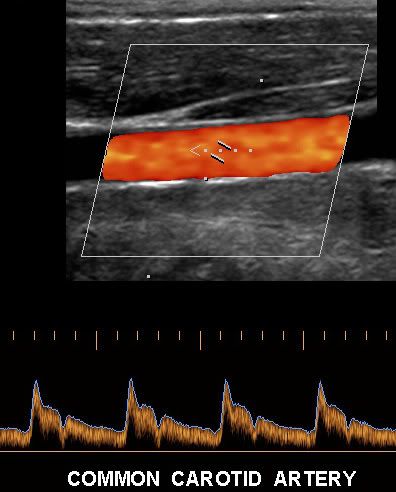
Use of carotid ultrasound to identify subclinical vascular disease and evaluate cardiovascular disease risk: a consensus statement from the American Society of Echocardiography Carotid Intima-Media Thickness Task Force
American Society of Echocardiography Carotid Intima-Media Thickness Task Force Journal of the American Society of Echocardiography 2008; 21 (2): 93 -111
There is great interest in identifying asymptomatic patients at high risk
who might be candidates for more intensive, evidence-based medical
interventions that reduce cardiovascular disease (CVD) risk. Measurement
of carotid intima-media thickness (CIMT) with B-mode
ultrasound is a noninvasive, sensitive, and reproducible technique for
identifying and quantifying subclinical vascular disease and for evaluating
CVD risk. To address issues of standardization and help
improve the availability of experienced clinical laboratories that can
perform high-quality CIMT studies, this consensus document provides
recommendations for the use of carotid ultrasound for identifying
and quantifying subclinical vascular disease and for evaluating
CVD risk in clinical practice. Nine published prospective studies that
included at least 1000 asymptomatic participants have examined
CIMT and CVD risk. Each study demonstrated that CIMT was
significantly associated with risk for myocardial infarction, stroke,
death from coronary heart disease, or a combination of these events.
In most of these studies, the ability of CIMT to predict future CVD
events was independent of traditional risk factors. Furthermore, 9
large studies have demonstrated similar or greater predictive power
http://rapidshare.com/files/95600633/Carotid_Ultrasound.pdf.html
Emergency Ultrasound, 2002

Number Of Pages: 435
Publication Date: 2002-10-24
Sales Rank: 437747
ISBN / ASIN: 0071374175
EAN: 9780071374170
Binding: Hardcover
Manufacturer: McGraw-Hill Professional
Studio: McGraw-Hill Professional
Average Rating: 4
Total Reviews: 1
Book Description:
A state-of-the-art reference on the training, techniques, and diagnostic
skills needed to perform successful ultrasound exams in the Emergency
Department. Includes detailed guidelines on performing ultrasound exams,
case studies, and side-by-side comparisons of normal and abnormal scans.
*
Length:39.4 MB
File:CHM
http://rapidshare.com/files/65878847...y.talented.rar<http://rapidshare.com/files/65878847/EmergUSG2002.by.talented.rar>
Search
QUICK CHAT

